Abdominal Anatomy
Most of the abdominal wall is muscular and extends between your thoracic rib cage and your bony pelvis (iliac crest). There are four important paired muscles in the anterior abdominal wall: three flat muscles (external oblique, internal oblique, and transversus abdominis) and one strap-like muscle (rectus abdominis).
 The External Oblique Muscle
This is the largest and most superficial of the three flat abdominal muscles.
Origin: external surfaces of 5th to 12th (lowest) ribs.
Insertion: linea alba, pubic tubercle and anterior half of the iliac crest.
The Internal Oblique Muscle
This is the intermediate layer of the three flat abdominal muscles.
Origin: thoracolumbar fascia, anterior two-thirds of iliac crest, and lateral half of inguinal ligament.
Insertion: inferior borders of 10th to 12th ribs, linea alba, and the pubic via the conjoint tendon.
The Transversus Abdominis Muscle
This is the innermost of the three flat abdominal muscles.
Origin: internal surfaces of 7th to 12th costal cartilage's, thoracolumbar fascia, iliac crest, and lateral third of inguinal ligament.
Insertion: linea alba with aponeurosis of internal oblique, pubic crest, and pecten pubis via conjoint tendon.
Actions of the Three Flat Abdominal Muscles
Unlike your rib cage, your abdominal wall is unsupported and unprotected by bone. The three-ply structure of its flat muscles are separated by a skin like sheet (aponeuroses) which joins each layer together thus increasing its strength, and stability. This provides considerable protection for your abdominal intestines, and other organs.
Acting separately, the flat abdominal muscles move the trunk. If the pelvis is fixed, both external oblique muscles can flex the trunk. Acting separately, one external oblique muscle can laterally flex the trunk and rotate it to the opposite side.
If the thorax if fixed, both external oblique muscles tilt the anterior part of the pelvis superiorly and flex the trunk.
Similarly, when the pelvis is fixed, one internal oblique muscle can flex the trunk and rotate it to the same side.
If the thorax is flexed, one internal oblique muscle can laterally flex the trunk and rotate the pelvis to the opposite side.
The Rectus Abdominis Muscle
This is a long, broad, strap-like muscle and is the principle vertical muscle of the front (anterior) abdominal wall.
Origin: pubic synthesis and pubic crest.
Insertion: base of your sternum - breast plate (xiphoid process)and 5th to 7th costal cartilages.
The two muscles are separated by a tendon sheath (linea alba). The lateral border of the rectus muscles and its sheath are convex and form a clinical important surface marking, known as the linea semilunaris.
Most of the rectus abdominis muscle is enclosed in the rectus sheath formed by the skin like layer (aponeuroses) of the three flat abdominal muscles.
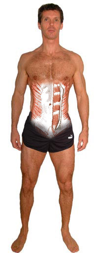
The anterior layer of the rectus sheath is firmly attached to the rectus muscle at three or more tendinous intersections. When this muscle is tensed in muscular persons, each stretch of muscle between the tendinous intersections is indicated by grooves in the skin between the muscle bulges - your six-pack.
They are usually located at the level of the xiphoid process, umbilicus, and halfway between this structures. Joining your two sides of the rectus abdominis muscle is a tendinous sheath called "The Linea Alba", which gives the appearance of a visible vertical line between your abdominal muscles.
Actions of the Rectus Abdominis Muscles
In addition to helping the other abdominal muscles to compress the abdominal organs, these muscles depress the ribs and stabilize the pelvis during upright movement. The fixation of the pelvis enables the thigh muscles to act effectively.
During lower limb lifts from the supine position, the rectus abdominis muscles contract to prevent tilting of the pelvis by the weight of the limbs.
Posterior Abdominal Wall
The posterior abdominal wall is composed principally of muscles and fascia attached to the vertebrae, hip bones, and ribs.
There are three paired muscles in the posterior abdominal wall that are clinically important: psoas major, iliacus, and quadratus lumborum.
Psoas Major is a thick, powerful muscle that passes from the abdomen to the inside of your the top of your thigh and is your strongest flexor (lifting your leg) muscle at the hip joint.
The iliopsoas muscles help flex the trunk, as in raising your upper body during a normal sit-up.
The quadratus lumborum extends and laterally flexes the vertebral column, and fixes the 12th rib during inspiration.