Physiology
The importance of the role of the musculoskeletal system.
When we talk about stretching the body, we are describing stretching a system known as the musculoskeletal system. This system includes the bones of the skeleton, along with the ligaments, muscles and their tendons, which help to move and support these bones.
The musculoskeletal system has developed primarily for the purpose of locomotion or movement. It also provides an excellent supporting/protective system to the vulnerable internal organs, whose main role is to provide the musculoskeletal system with nourishment, stimulation, and purification. Many people consider the nervous system, including the brain; or the circulatory system, including the heart, to be the most important systems. It goes without saying that if the heart stops and the brain ceases to function, the body dies. However, without the body being able to move and generate energy and heat, search for food, help circulate the blood and lymph, expand the rib cage in order to breathe, and perform numerous evasive movements in order to protect itself, it would not be able to exist and function naturally. Therefore, it would be quite reasonable to state that the musculoskeletal system is the most important system. By accepting the importance of its role of importance, we must also recognize that the need to maintain it in top condition should be paramount. If this is difficult to understand and accept, consider the plights of people who suffer from advanced muscular dystrophy, or are totally paralyzed. Gradually their organs fail, their lungs cannot function properly, or unaided, due to weakness in the respiratory muscles, and the lack of posture causes a stasis in both the lungs and the circulation, normally resulting in severe infections and eventual death.
The components of the musculoskeletal system.
There are three main components to this intricate system, all working in conjunction with each other, and all having some effect on the system as a whole. The first component is the skeleton. This consists of numerous bones, whose shape and structure reflects their role, and which are brought into contact with each other to make a “joint”. This gives rise to a structure made of a very solid, strong material, which is able to articulate. The outer layers of the bones consist of a hard, dense material, while the centers are made of a spongy cortex that makes the bones lighter, otherwise we would not be able to move our own weight! This cortex also provides a medium for the rich network of blood vessels to penetrate through the bones, providing nourishment, and transporting away the new red blood cells that are manufactured in some bones. The ends of all articulating bones are covered in a cartilaginous surface (hyaline), which helps to protect the bones against the wear and tear caused by continual movement and friction, and allows the bones to “glide” over, or against, each other. Articulating joints are called synovial joints, because, as well as the bone-ends being covered with cartilage, the whole joint is encased in a capsule, a bit like a sleeve/tube enveloping both ends together. Lining this capsule is a membrane called the synovial membrane, which produces a fluid (synovial fluid) that lubricates the surfaces.
To keep these joints aligned properly with one another is our second component, the ligaments. These are strap-like structures consisting of dense fibrous tissue, which are positioned in such a way as to allow movement, but also to restrict the range of movement to within a safe level to prevent damage to adjacent tissues and structures. Ligaments also assist in support. Ligaments are normally positioned around the joint and outside of the joint capsule (extracapsular).
If you have ever strained your ankle or knee, you will find this easy to understand. The ligaments that support these two areas allow flexion, extension, and some degree of rotation. In the case of the knee there are two important extra ligaments called “cruciates” (because they cross over each other). These are located inside the joint capsule (intracapsular), and limit the forward/backward shift of the femur (thigh bone) on the tibia (leg bone). Damage to any of these ligaments, as a result of injury and trauma, can give rise to sudden misalignment of the bones, pain and swelling in the joint itself, and instability, which frequently leads to subsequent recurrence of the same injury. Treatment is normally based on restabilizing the joint with some form of strapping, and because this normally works well, demonstrates the importance of the role of the ligaments in both posture and joint stability. It is also important in a newly strained joint that you reduce the swelling that usually occurs as a result of inflammation. If the swelling, which is a build-up of fluid in the tissues, is allowed to persist, it will cause some structures to be stretched, especially unaffected ligaments, thus aggravating the problem. Reducing the inflammation by locally applied cold compresses and/anti-inflammatory gel will greatly assist the overall healing process. A homeopathic remedy called Arnica is also very beneficial and is available in tablet or cream form. It helps to promote healing in bruised tissue, a symptom that is often not immediately obvious in strained or damaged joints. All of these remedies are most effective if initiated immediately after the injury occurs.
Our third component is the muscles-probably the most complex yet versatile group of the three. Without muscles there can be no movement. Each muscle has a main part, or “belly”, and two ends called tendons. Muscles are normally firmly attached on the two bones either side of a joint via their tendons, and when the belly contracts, it shortens, and in doing so the tendons pull against their attachment on the bones shortening the distance between the two bones, and causing a movement at the joint. Skeletal muscles can also contract to cause other types of movement not involving joints, e.g., facial muscles contract to produce a smile; the diaphragm contracts causing the lungs to inflate. Skeletal muscle is made up of thousands of muscle fibers all joined together in bundles and separated by layers of fibrous connective tissue. Many of the layers of fibrous tissue extend the whole length of the muscle, and end as part of the tendon that will attach to the surface of the bone. This continuity helps to provide the strength to the muscle structure. The shape of a muscle and the size, shape, and direction of the muscle fibers, can vary greatly between one muscle and the next, as they are all are designed to fulfil a particular role. The saying “structure is related to function” is most applicable here. Muscle and bone are two completely different tissues, therefore they need a third substance to help bond them together, just like wood and glass need putty. This third tissue is called periosteum, it is an outer covering to the bone, which is fibrous, allowing the fibers of the muscle tendon to blend with it, and thus form an attachment between tendon and bone.
The condition known as tennis elbow arises as a result of too much tension being exerted by the tendon at its periosteal site, just above the elbow. The site at which the tendon attaches can be quite small in area, yet the pull on that area can be extremely high..
With persistently tight extensor forearm muscles, the continual pull by the extensor tendons can actually cause the beginning of a separation of the various fibrous layers of the periosteum. This leads to inflammatory changes taking place within the area, which in turn leads to further irritation of the muscle tendon, and as such a vicious circle is set up. To treat this type of strain, you need to reduce both the tension within the muscle by rest and/or stretching, and reduce the inflammation locally with icepacks, anti-inflammatory gel, or occasionally a cortisone injection is required.
How do muscles work?
Skeletal muscle tissue is connected to both the nervous system and the circulatory system, and needs both in order to function.
The nerve supply:
First, a muscle cannot initiate a contraction without a nerve impulse stimulating an electrical impulse at a site known as the neuromuscular junction. This simply translates as “no nerve impulse, no muscle activity,” which is clearly demonstrated where nerves have been severed or damaged due to trauma and where paralysis occurs. Where each nerve cell sends an impulse to a muscle cell a “motor unit” is formed. Depending on how many muscle fibers are supplied by that motor unit will determine the type of function of that particular muscle. If the motor unit is linked to just a few fibers (taking into account that this particular muscle may consist of thousands of motor unit/fiber components), a refined contractile movement of part of the muscle can be obtained, giving a more precise movement. This is seen in the muscles of the hands and face, for example. In muscles where one motor unit is linked to thousands of fibers, a more basic substantial contraction is involved, e.g., in the large bulky muscles of the legs.
The stimulation by the nerve impulse releases two main chemical components-calcium and a high-energy compound called adenosine triphosphate, or ATP. In simple terms the calcium “switches on” the muscle and the ATP provides the energy for the contraction activity to be performed (this chemical release activates extremely tiny filaments that make up the muscle fiber, to slide across each other, causing a shortening of the muscle fiber, which results in a contraction). The withdrawal of calcium back into the cells “switches off” the muscle contraction and “switches on” muscle relaxation. The strength of the muscle contraction is influenced by many factors. These include the initial length of the muscle fibers, the metabolic state of the fiber, the number of motor units and fibers activated, and the load put on them.
The stretch receptors:
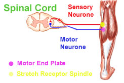 The nervous system supplies muscles with the means to contract (motor), but it also provides it with a second system, which gives feedback (sensory) to the brain, telling it what is going on within the muscle and where. This sensory system includes specialized sensory (stretch) receptors, called “muscle spindles”, and “Golgi tendon organs”, which are capable of detecting the degree of stretch in both the muscle, and its junction with the tendon, respectively. These are very important receptors, as they act as warning systems and will prevent the muscle from damage due to overstretch: as the muscle is stretched the muscle spindles are stretched too. When this happens, the stretch receptors are activated sending a signal to the brain, which initiates a motor impulse to encourage contraction to occur, thus preventing any overstretching of the fibers. This feedback mechanism is called “the reflex arc”.
In summary, the motor nerve fibers instigate an impulse that leads to a contraction, which results in the stretching of other muscles. Stretching sends off a signal to the brain, which then leads to the stretched muscle contracting and regaining its shape/length, which is important in protecting it from injury. The signals that are sent from the muscle spindle to the brain are fired at a particular rate, which helps to clarify to the brain the exact degree of stretch, i.e., the change in length and the speed of change. In a slow, controlled stretch it is possible to re-train these receptors to become accustomed to a new length, before they trigger off the impulse to contract to protect themselves.
Skeletal muscles almost always act in groups, and movements are produced by the coordinated action of several muscles. The way these groups work will vary depending on their role. Having stated that if stretch occurs an automatic signal will be activated to cause a contraction and end the stretch, it is necessary to add that this is not always so. There are some muscle groups that work in cooperation with each other as agonists and antagonists, e.g., the biceps and the triceps. As the agonists contract, the antagonists are forced to relax. If this did not happen you would never be able to bend your arm, as there would be an ongoing power struggle between the two sets of muscles. The muscles, or muscle group, that actually cause the movement, are normally referred to as the “prime movers”. Muscles that contract at the same time as the prime mover and help to stabilize a part of that movement, or reinforce it, are known as “synergists”. An example of this is in the calf where the gastrocnemius muscle is the prime mover and soleus, which is located underneath the gastrocnemius, is the synergist, The soleus muscle is important in maintaining posture.
Continuous feedback occurs automatically between muscle tissue and the brain, via the nervous system. If damage or disease occurs to the peripheral nerve itself, or at the neuromuscular junction, complete loss of tone can result. Causes can range from trauma or pressure, but also include disorders such as diabetes, multiple sclerosis and the various forms of muscular dystrophy. Damage to the upper motor neurone within the brain usually results in disorders of increased tone and spasm.
The blood supply to the muscles
The circulatory system not only supplies blood by means of its arterial system, but also allows blood to drain from the tissues via its venous system. In the case of muscle tissue both systems are equally important.
The arterial supply to the muscle is essential in providing the muscle with a continuously available source of calcium and the components to needed to manufacture ATP within the cells. The venous drainage allows all the waste by-products of muscle activity to be taken away from the muscle itself. One of the main by-products is lactic acid, and a build-up of this within the muscle is what can cause what we commonly call “a stitch”.
Taking into account these facts should help you understand the overall concept of keeping the body supple and flexible, as the circulation, particularly the venous drainage element, requires rhythmic movement to encourage blood flow back toward the heart and lungs for purification. Extremely tight calf muscles can prevent this from happening satisfactorily. The regular rhythmic contractions of both the calf and buttock muscles combine to form a “pump” mechanism, which, in conjunction with a valve mechanism in the main veins of the lower limbs, encourages blood flow upward against gravity. People who sit all day or stand still in one place for long periods of time can compromise this mechanism. Typical symptoms of this are tired and achy feet and legs, and swollen feet and/or ankles. The ache directly results from the waste products being held too long within the muscle tissue.
Posture and muscle tone
Gravity has a continual pull on the various part of the body at all times. Our evolutionary development, ending with us standing upright on two legs, has meant extensive adaptation of our mechanism over the years. There is a noticeable difference in the human spinal muscle tone and development compared to that of four-legged mammals. These gradual changes have led to a redistribution of muscle bulk and fat around the body as a whole. Posture has to be maintained using the minimum amount of energy, which is why people with poor posture suffer with tired aching muscles. This may sound too simplistic, but any posture, whether it is sitting or standing, bending or stretching, is achieved by the combination and coordination of different muscle groups contracting and relaxing in order to achieve balance. If one or more groups are not able to take advantage of regular episodes of relaxation in between their role as contractors (perhaps because in doing so balance would be lost), then these groups will eventually fatigue, having used excessive energy to maintain their workload, and their tissue state would suffer. In order to consider good posture; look at yourself sideways on in the mirror sideways and ask yourself “Does more stick out the front than it does the back?” Sounds crazy? It shouldn’t, because if your body is being encouraged to lean forward due to poor abdominal tone, excess weight, or slumped and rounded shoulders, what do you think is stopping you falling flat on your face? The answer is overworked, tight, and tired back and calf muscles. This is why it is important to keep you weight down, maintain good abdominal tone, and help to keep those back muscles loose and relaxed. Which is the reason that regular stretching of your back and lower limb muscles are really important. Statistics show that more days are lost at work from back strain than any other injury.
Age-related problems and changes due to injury
Skeletal muscle undergoes various methods of repair. In young healthy adults with a steady supply of the components essential for these repairs, damaged tissue will be repaired as new, resulting in continuity of elastic muscle fibers. In older people, as fibers degenerate naturally or become damaged, they are not repaired with elastin (a protein) containing components, but are instead replaced with fibrous tissue, a process called fibrosis. This fibrous connective tissue may be relatively strong, but is unable to stretch as well as muscle, and, because it contains no actual muscle fibers, cannot contract. In essence, a muscle can gradually become so fibrosed, that the ratio of muscle fibers to connective tissue becomes so low that the muscle loses its ability to function as a muscle and weakness occurs. While this is an unfortunate part of the ageing process, a similar situation can occur as a result of repeated injuries at the same site. This is often seen in footballers, where the calf muscles can receive severe injuries from kicks. The tissues heal with fibrotic changes, which can lead to an inability to stretch the calf properly, and can subsequently lead to injuries occurring to other parts of the body as a result of an inability to move freely. Fibrotic tissue in the calves does not possess the same amount of elasticity and therefore cannot assist the “calf pump mechanism”. People who undergo surgery for repair to damaged tissue should aim to begin stretching the area gently, yet persistently, to prevent connective tissue layers sticking together as adhesions and giving the same long-term problems as fibrosed muscles.
In summary, throughout our lives we should make regular effort to maintain our body by stretching, keeping our joints supple, and getting our circulation moving.